 Micrograph showing extreme nuclear atypia in cancer (glioblastoma). Brain biopsy. HPS stain.
 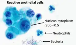 Cytopathology of reactive urothelial changes, Pap stain, showing urothelial cells with enlarged nuclei but a nucleus-cytoplasm ratio of less than 0.5. There are bacteria, as well as an inflammatory response of neutrophils, providing a cause for the changes.
 Nuclear atypia refers to abnormal appearance of cell nuclei. It is a term used in cytopathology and histopathology. Atypical nuclei are often pleomorphic.
Nuclear atypia can be seen in reactive changes, pre-neoplastic changes and malignancy. Severe nuclear atypia is, in most cases, considered an indicator of malignancy.
See also